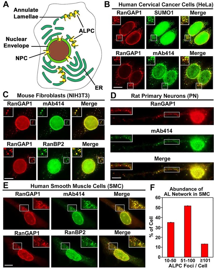
Fig 1. SUMO1-modified RanGAP1 localizes to both NPCs and ALPCs in a variety of mammalian cells.
(A) The diagram shows that compared to NPCs in the nuclear envelope, ALPCs are embedded in the membrane cisternae of annulate lamellae that are often connected to the membrane network of ER. (B) Human cervical cancer cells (HeLa) were double-labeled with anti-RanGAP1 antibody and anti-SUMO1 mAb (21C7) or mAb414 for staining NPCs and ALPCs and then analyzed by immunofluorescence microscopy. (C) Mouse embryonic fibroblasts (NIH3T3) were double-stained with anti-RanGAP1 antibody and mAb414 or anti-RanBP2 mAb. (D) Rat primary cortical/hippocampal neurons (PN) were double-labeled with anti-RanGAP1 antibody and mAb414. (E) Human bronchial/tracheal smooth muscle cells (SMC) cells were double-stained with anti-RanGAP1 antibody and mAb414 or anti-RanBP2 mAb. Bar, 10 μm. The boxes at the top corner of each image show an enlarged version of inlets. (F) Annulate lamellae are highly abundant in SMC cells. 60 SMC cells were double-stained with anti-RanGAP1 antibody and mAb414. All the ALPC foci in each cell were counted under Olympus inverted IX81 fluorescence microscope using Z-stacks. The number of ALPC foci per cell was classified into three categories (10–50, 50–100 and ≥100), and the percentage of cells in each category was indicated. Each column represents the mean value ± SEM (N = 60) (ALPC foci/cell: 10–182; Average = 63).

