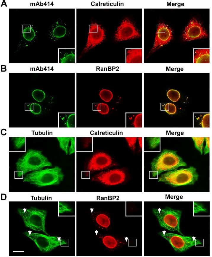
Fig 4. The ALPC-associated RanBP2/RanGAP1*SUMO1/Ubc9 complexes are distributed within the network of ER but not the tips of cell extensions.
(A-D) HeLa cells were double stained with mAb414 and calreticulin antibody for labeling ER network (A), mAb414 and RanBP2 antibody (B), tubulin and calreticulin antibodies (C), and tubulin and RanBP2 antibodies (D) followed by immunofluorescence microscopy. The enlarged versions of inlets are shown at the bottom or top corner of each image (A-D). The arrows indicate the positions of the ALPC-associated RanBP2/RanGAP1*SUMO1/Ubc9 complexes that are most distant from the corresponding nucleus (D). The immunofluorescent images were taken using Olympus inverted IX81 widefield fluorescence microscope with U-Plan S-Apo 60×/1.35 NA oil immersion objective. Bar, 10 μm.

