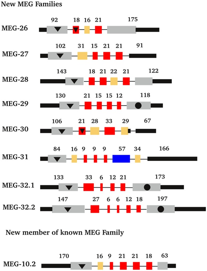
Fig 5. Exon structure of newly designated MEG genes.

Exons are colour coded as follows: red, symmetrical micro-exons; grey, coding region of the flanking exons; black, non-coding regions; yellow, non-symmetrical micro exons; purple, regular sized symmetrical exons. Other features: black circle, exons that code for transmembrane anchors; black triangle, exons that code for signal peptides. In the case of MEGs 26 and 30 part of the signal peptide is coded by the first micro-exon in addition to the terminal exon.
