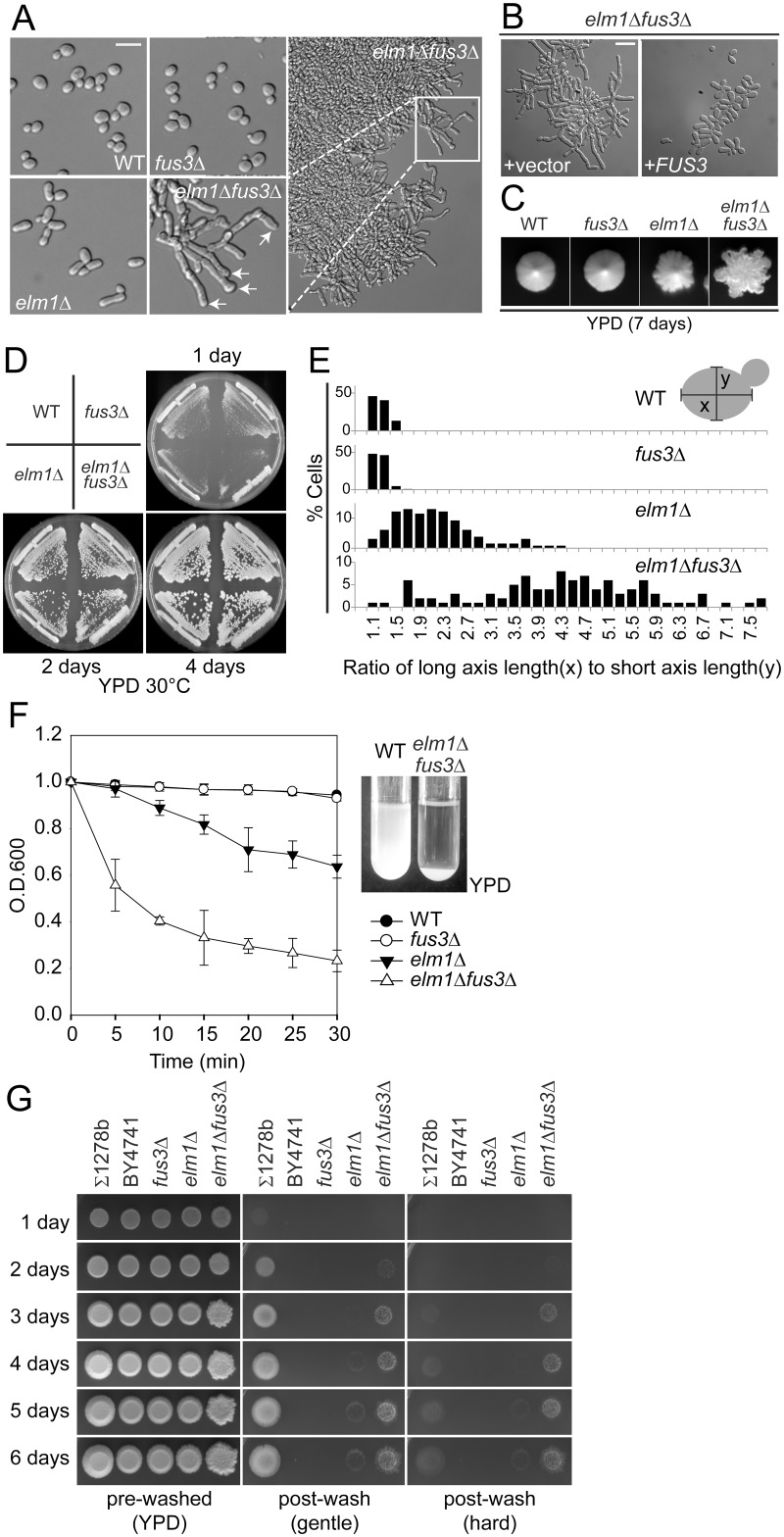
Fig 1. Filamentous growth of elm1Δ fus3Δ cells.
All of indicated strains (MY8092, 12158, 12886 and 12948) were BY4741-derivative haploids and grown to exponential phase in liquid YPD at 30°C unless stated otherwise. (A) Arrows indicate the buds that emerge from the pole opposite previous division site. (B) elm1Δ fus3Δ (MY12948) harboring empty vector (MR1865) or pCEN-FUS3 (MR5048) were grown in liquid synthetic complete (SC) medium containing 2% glucose. (C) Cells were streaked for single cells on YPD plate at 30°C for 7 days, and representatives in a zone of low colony density were photographed. (D) Indicated cells (MY8092, 12158, 12886 and 12948) were streaked on YPD solid medium and incubated at 30°C. (E) The ratio of long axis (x) to short axis length (y) was expressed in a histogram. For example, 1.1 and 1.3 at the x-axis indicate the range from 1.00 to 1.19 and 1.20 to 1.39, respectively. (F) Flocculation assay was performed as described in Materials and methods. Data are representative of three independent experiments and expressed relative to WT. (G) Cells were spotted on YPD plate and grown at 30°C for 6 days. Haploid Σ1278b (MY13465) was used as a positive control of invasive growth. (A and B) Bar, 10 μm.

