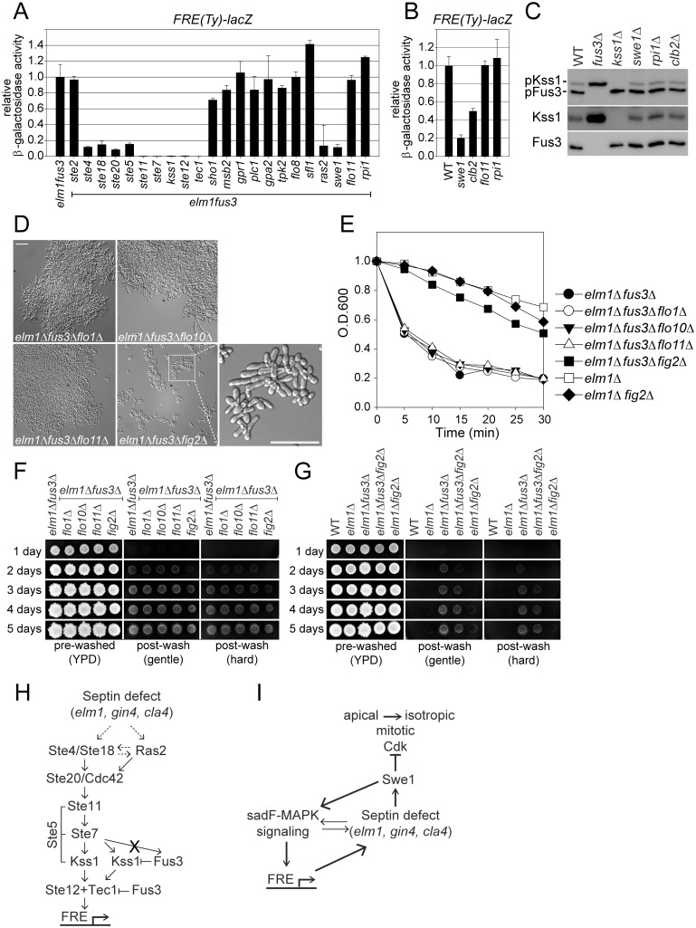
Fig 6. Identification of sadF pseudohyphal signaling pathway.
All of strains were BY474-derivative haploids and grown to exponential phase in liquid YPD at 30°C unless stated otherwise. (A and B) Indicated strains (MY8092, 12948, 13122, 13125, 13145, 13281, 13313, 13315, 13317, 13319, 13378, 13380, 13382, 13384, 13413, 13417, 13500, 13502, 13504, 13506, 13508, 13531, 13582, 14331, 14332, 14333, 14334 and 14335) harboring FRE(Ty)-lacZ (MR6857) were used. β-galactosidase activity was determined as described in Fig 5C. Data are expressed relative to (A) elm1Δ fus3Δ or (B) WT. Data are mean ± standard deviation from three independent experiments. Note that (A) elm1Δ fus3Δ, elm1Δ fus3Δ kss1Δ, elm1Δ fus3Δ tec1Δ and (B) WT are identical to Fig 5C. (C) MY8092, 12886, 13144 and 13145 strains were used. Each protein was detected as described as Fig 5G. (D-G) Indicated strains (MY12158, 12948, 13413, 13901, 13903, 13907 and 14336) were used. (D) Bar, 30μm. (E) Flocculation assay was performed as described in Fig 1F. Data are representative of three independent experiments and expressed relative to WT. Note that elm1Δ and elm1Δ fus3Δare identical to Fig 1F. (F and G) Plate washing assay was performed as described in Fig 1G. (H and I) A model for sadF pseudohyphal signaling pathway. See the Discussion for details.

