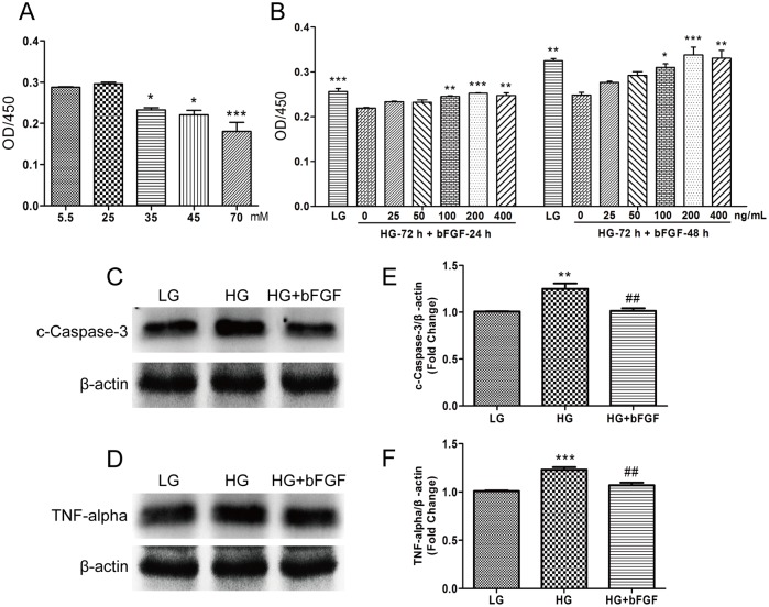
Fig 2. bFGF protects VECs against the deleterious effects of HG on VEC viability and proliferation.
(A) Cell proliferation after 72 hours of HG treatment (25–70 mM), and (B) proliferation of cells treated with different concentrations of bFGF for 24 or 48 hours following 72 hours of HG treatment, measured by CCK-8 assay. (A) and (B) data represent mean values ± SE of four independent experiments (*P < 0.05, **P < 0.01, ***P < 0.001). Immunoblotting analysis for detection of cleaved Caspase-3 (C) and TNF-α (D) in VECs treated with HG (35 mM) for 72 hours, and then incubated with 100 ng/mL bFGF for 1 hour. LG: 5.5 mM glucose in culture medium. Signal intensities for cleaved Caspase-3 (E) and TNF-α (F) were normalized to that of β-actin. Results are presented as fold changes relative to VECs grown in medium containing 5.5 mM glucose (LG). (E) and (F) Data represent mean values ± SE of four independent experiments, relative to the LG group (**P < 0.01, ***P < 0.001) and the HG group (##P < 0.01).

