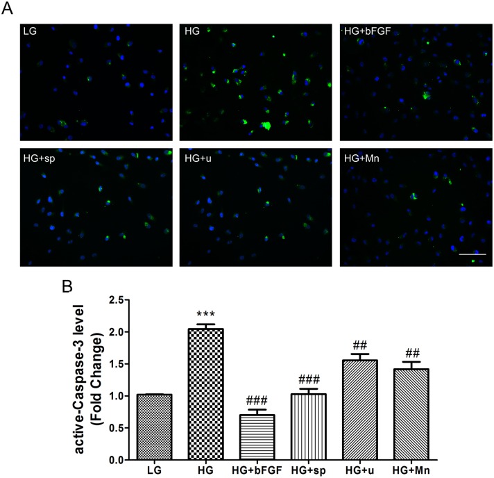
Fig 7. High-glucose induces apoptosis in VEC.
(A) Apoptotic responses in VEC were analyzed by detection of active-Caspase-3 immunofluorescence using a-Caspase-3 antibody. Cells were treated with low glucose (LG) or high glucose (HG) for 72 hours before treated with 100 ng/mL bFGF, 1 μM sp600125 (sp), or 1 μM U0126 (U) or 10 μM MnTmPyP for 1 hour. Bar = 100 μm. (B) Fluorescence levels in ten different cells were measured using the Image Pro Plus software. Data represent mean values ± SE of four replicates, relative to the LG group (***P < 0.001) and relative to the HG group (##P < 0.01, ###P < 0.001). DAPI was used to stain nucleus. Green and blue signals indicate a-Caspase-3 and nucleus respectively.

