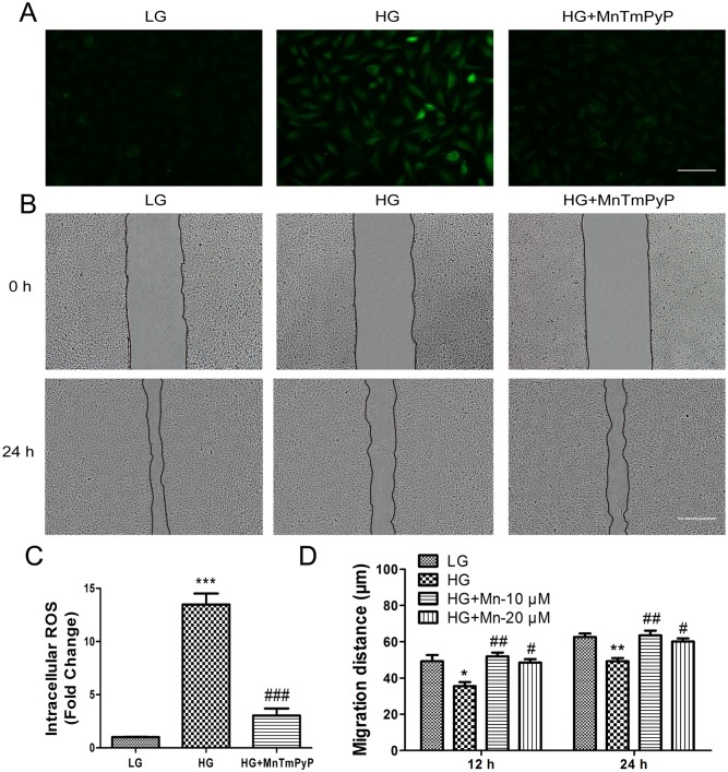
Fig 8. High glucose-induced accumulation of intracellular ROS levels delays VEC migration.
(A) Cells were pretreated with high glucose (35 mM) for 72 hours, and then incubated with the ROS scavenger MnTmPyP (10 μM) for 1 hour. Intracellular ROS levels were measured using DCFH-DA dye. Bar = 100 μm. (B) Wound scratch assay was performed to analyze the effects of 10 μM MnTmPyP in human VECs. LG: medium containing 5.5 mM glucose. Bar = 500 μm. All experiments were performed after exposure of cells to 5 mg/mL mitomycin C (to inhibit proliferation) for 1 day. (C) Fluorescence levels in ten different cells were measured using the Image Pro Plus software. Data represent mean values ± SE of four replicates, relative to the LG group (***P < 0.001) and relative to the HG group (###P < 0.001). (D) Cell migration distance was determined according to the data shown in (B). Data represent mean values ± SE of ten replicates, relative to the HG group (*P < 0.05, **P < 0.01).

