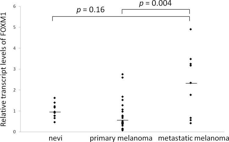
Fig 2. The results of the quantitative RT-PCR analyses of melanoma and nevus tissue samples.

The FOXM1 expression (normalized to GAPDH) in the patients with primary melanoma (n = 25), metastatic melanoma (n = 9) and nevi (n = 10) is shown. The bars indicate the median values.
