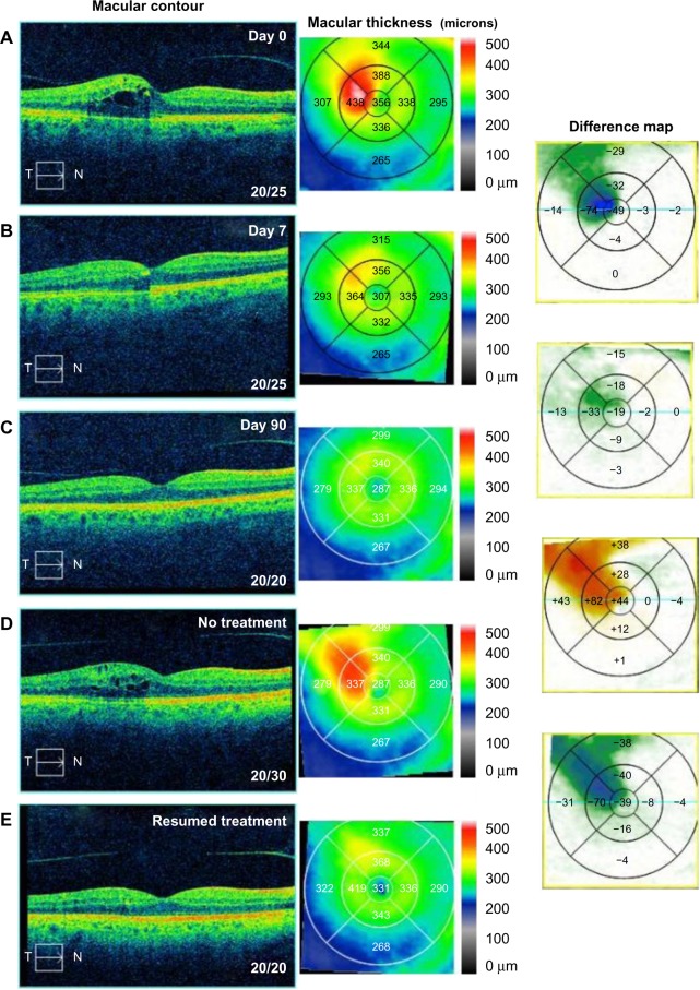
Figure 6.
Optical coherence tomography studies for case 6 (branch retinal vein occlusion).
Notes: (A) Macular contour and thickness at presentation. (B and C) Macular contour and thickness and macular change analysis (B) 7 days and (C) 90 days following treatment. (D) Increased cystoid macular edema with cessation of treatment and (E) improvement with resumption of treatment.
Abbreviations: T, temporal; N, nasal.

