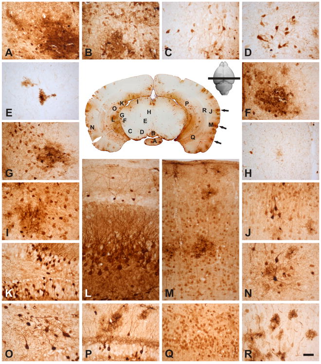
Fig. 5.
rAAVrh10 injected into the lumbar cistern transduced regions in the cortex, hippocampus and rostral midbrain: (A) medial mammillary nucleus, (B) rostral interpeduncular nucleus. (C) substantia nigra, (D) parabrachial pigmented nucleus, (E) red nucleus, (F) posterior intralaminar thalamic nucleus, (G) medial geniculate nucleus, (H) periaqueductal gray, (I) superior colliculus, (K, O, P) dentate gyrus, (L, Q) CA3 of the hippocampus, (J, M, N, R) cortex. Note that the temporal cortex had higher level of transduction compared with the parietal cortex; in hippocampus, the most prominent level of transduction was found in dentate gyrus and CA3; in the midbrain, most transduction was in the peripheral regions. Arrows in the central panel point to patches of transduction that appeared to project from the edge into the parenchyma of the brain (see text). Bar = 50μm.

