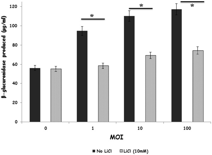
Figure 4.
Inhibition of GSK-3β regulates primary degranulation by avian heterophils stimulated with S. Enteritidis. Heterophils were pre-incubated with LiCl for 1 h and then stimulated with various MOI of S. Enteritidis for 1 h at 41°C. Heterophils (8 × 10⁶) were incubated with either RPMI 1640 medium alone or the GSK inhibitor, LiCl at room temperature for 1 h. The heterophils were then stimulated with various MOI of S. Enteritidis (1, 10, or 100) for 1 h at 41°C. The reaction was stopped by transferring the tubes containing the cells to an ice bath for 5–10 min. The cells were then centrifuged at 250 g for 10 min at 4°C. The supernatants were then removed and used for the assay. A 25 μl aliquot of each supernatant was added to quadruplicate wells in a non-treated, black CoStar flat-bottom ELISA plate and incubated with 50 μl of freshly prepared substrate (10 mM 4-methylumbelliferyl-β-D-glucuronide, 0.1% Triton X-100 in 0.1M sodium acetate buffer) for 4 h at 41°C. The reaction was stopped by adding 200 μl of stop solution (0.05M glycine and 5 mM EDTA; pH 10.4) to each well. Liberated 4-methylumbelliferone was measured fluorimetrically (excitation wavelength of 355 nm and an emission wavelength of 460 nm) with a GENios Plus Fluorescence Microplate Reader (TECAN US Inc., Research Triangle Park, NC, USA). These values were converted to micromoles of 4-methylumbelliferone generated using a standard curve of known concentrations. *p ≤ 0.01.

