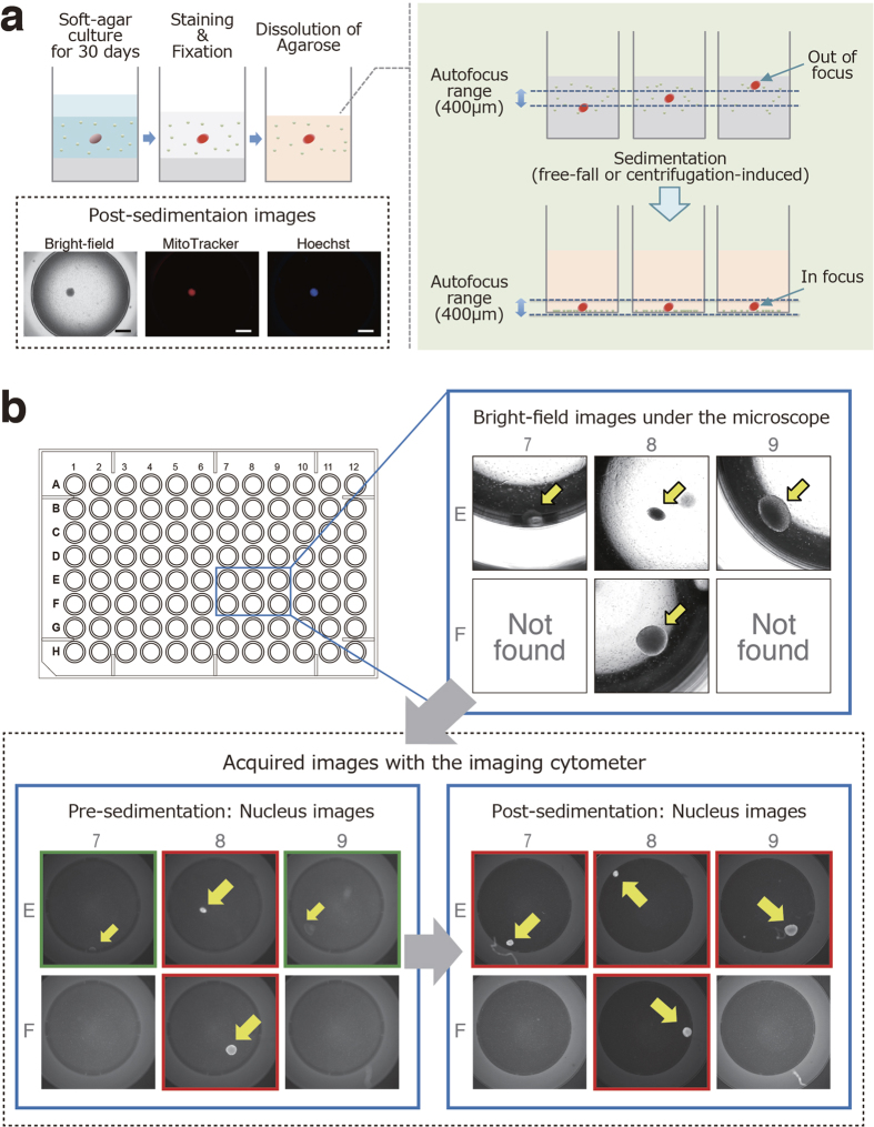
Figure 3. High-precision 2D imaging of colonies in soft agar culture using the imaging cytometer.
(a) Although the imaging cytometer has the ability to autofocus on colonies formed in the soft agar media, it cannot actually recognize colonies out of the autofocus range (400 μm) of the equipped lens. To overcome difficulties in focusing on colonies in soft agar media with the imaging cytometer, the sedimentation of the floating colonies in the soft agar media into the bottom of the wells was attempted by dissolving the agarose. Representative post-sedimentation images are shown (magnification, 20×; scale bars, 1 mm). (b) Cell suspensions containing one HeLa cell and 12,500 hMSCs per well of 96-well plates were cultured in soft agar media for 30 days. Representative bright-field images of formed colonies with a light microscope are shown (upper right). These colonies were stained, fixed and subjected to high-content screening. Although some fluorescent images of colonies were not captured due to failed lens focus (green-edged images in the lower left panel), the sedimentation of the colonies enabled the imaging cytometer to clearly capture images of the colonies (red-edged images in the lower right panel). hMSCs, human mesenchymal stem cells.

