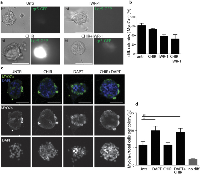
Figure 3.
Representative images of OC spheres at day 5 in vitro cultured in presence of CHIR99021 (10 μM), IWR-1 (2.5 μM) or the combination of the two. Brightfield (left) and GFP (right) channels are shown. Scale bar 50 μm. (b) Quantification of the fraction of colonies containing MyoVIIa + cells over the total colonies counted (n = 20 from 2 independent experiments) after 15 days of differentiation. CHIR99021 and IWR-1 are added exclusively during the first 5 days in sphere culture. (c) Representative examples of spheres after differentiation immunostained for MyoVIIa (green) and DAPI (blue). Cells are treated with/without CHIR99021 in sphere cultures and further plated for differentiation with/without DAPT (1 μM) for additional 15 days. Maximum intensity Z projections of confocal stacks are shown as merged and single channels. Scale bar 50 μm. (d) Quantification of the percentage of MyoVIIa positive cells per colony. Cells are treated with/without CHIR99021 in sphere culture and further plated for differentiation with/without DAPT (1 μM) for additional 15 days. Mean +/− SEM *p < 0.05 (Kruskal-Wallis test with Dunn’s correction). 70–80 spheres are counted per condition and derived from 7 independent experiments. Untreated cells (primary spheres) at day 5 in vitro are plated in parallel and after 1 day fixed for immunostaining. MyoVIIa + cells are counted prior to differentiation to estimate carried over hair cells from the isolation (gray bar). All samples are statistically significantly different from negative control. (Kruskal-Wallis test with Dunn’s correction).

