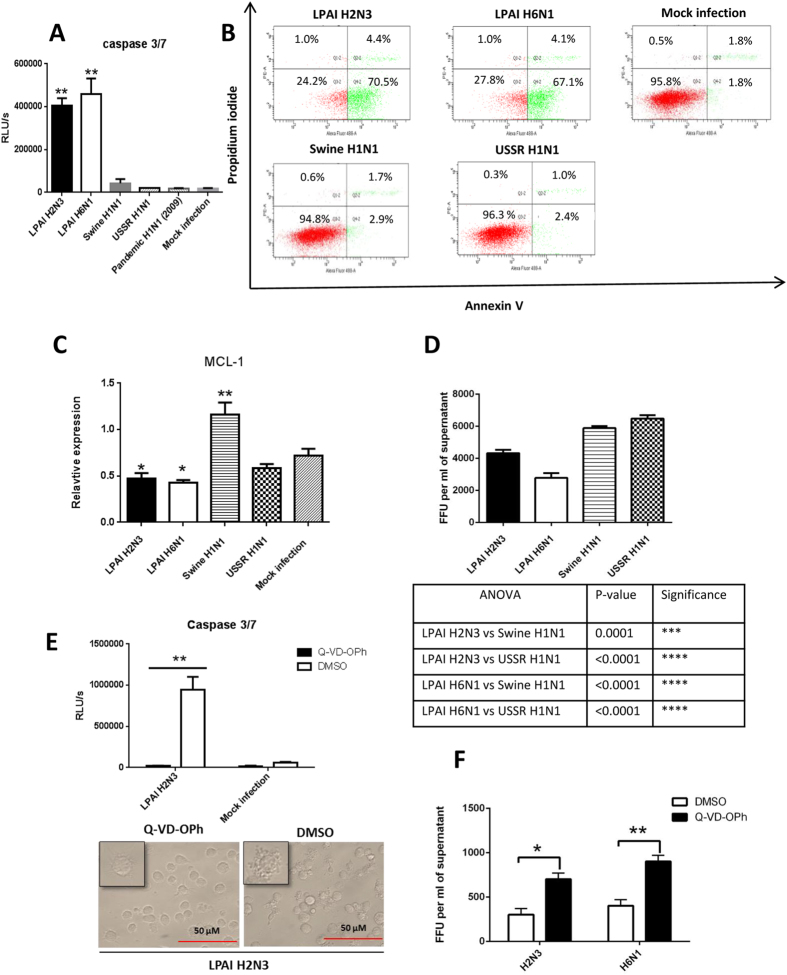
Figure 3. Avian influenza virus infected PAM displayed rapid apoptosis and reduced progeny virus release.
PAM were infected with avian and mammalian viruses as indicated at MOI of 1.0 for 6 h to determine host responses in (A) caspase 3/7 activation, (B) relative presentation of annexin V and uptake of propidium iodide (PI), (C) messenger RNA expression of Mcl1, and (D) infectious virus release into culture supernatants. PAM infected with avian influenza viruses showed (A) much higher caspase3/7 activation, and (B) more detectable annexin V with little evidence of necrosis as indicated by lack of PI uptake. (C) Marked apoptosis detected in avian virus infected PAM coincided with the down-regulation of Mcl1 (expression normalized to 18S rRNA) by avian H2N3 and H6N1 viruses. (D) Less avian than mammalian progeny virus was released from 6 h infected PAM, as determined by virus titration on MDCK cells with culture supernatants. (E,F) PAM were infected with LPAI H2N3 or LPAI H6N1 virus at 1.0 MOI; after 1 h of initial virus incubation the cells were washed with PBS and incubated in fresh infection culture media with 20 μM Q-VD-OPh, a pan-caspase inhibitor, for a further 5 h. (E) Infected PAM in the presence Q-VD-OPh showed no caspase 3/7 activation and no apparent cytopathic or apoptotic damage, unlike infected DMSO treated PAM. (F) Q-VD-OPh inhibition of apoptosis in PAM increased viable progeny release of H2N3 and H6N1 viruses. Data shown are the means of triplicate wells. Error bar = standard error of mean. *P < 0.05, **P < 0.001 (ANOVA with the use of Prism [GraphPad]).

