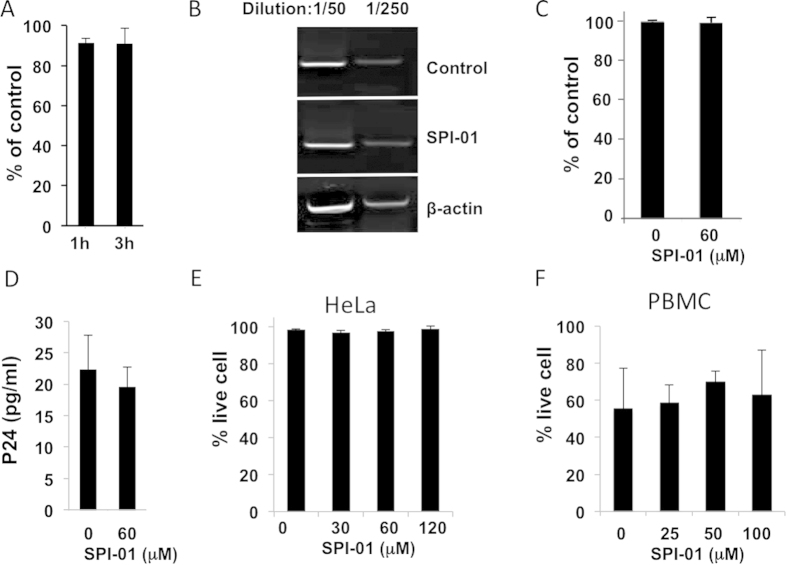
Figure 1. SPI-01 inhibited HIV-1 replication but did not inhibit production of virus.
(A) Expression of luciferase as percent of untreated control following transduction of HeLa cells with VSV-G pseudotyped pNL43LUCR-E- showing no significant inhibition by treatment of the cells with 60 μM SPI-01 for either 1 or 3 h. (B) SPI did not significantly block viral integration. HeLa cells treated overnight with 120 μM SPI-01, and transduced with DNase-treated VSV-G pseudotyped virus supernatant. To detect integration products, 48 h post infection the isolated total cellular DNA was analyzed by nested PCR using Alu- and viral-specific primers. β-actin primers were used for normalization. PCR products were diluted by 50 or 250 folds and visualized by ethidium bromide stained agarose gel showing the DNA bands after PCR. (C) Tat activation of HIV-1 LTR-luciferase after treatment with SPI-01. 293T cells were cotransfected with HIV-1-LTR-Luc and Tat expression plasmid and treated with 60 μM SPI-01. After transfection (24 h), luciferase activity was assessed, normalized against Renilla luciferase, and compared to the control to determine % activity. (D) VSV-G pseudotyped pNL43LUCR-E- viral production (assessed by p24 levels) in HeLa cells treated with 60 μM SPI-01 or mock treated. No significant reduction was observed. (E) Representative data showing that SPI-01 confers minimal toxicity. HeLa cells were treated with indicated concentrations of SPI-01 for 72 h. Dead cells were assayed by trypan blue staining. (F) CD8-depleted PBMCs, maintained in TCM 1000 ActiCyte media (BioE), challenged with HIV-1 NL4-3 (MOI of 0.01) were treated with SPI-01 at the indicated concentrations for 12 days. Percent of live cells were determined by the Live/Dead® Fixable Dead Cell Stain Kits (Life technologies).

