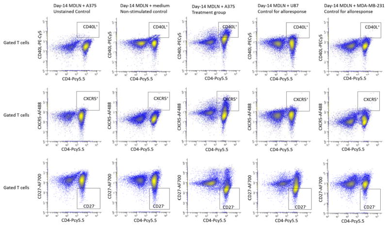
Figure 2.
FACS analysis of surface expression of CD40L, CXCR5 and CD27 by CD4+ cells in day-14 MDLN co-cultures with medium, A375, U87 and MDA-MB-231. Various MDLN co-cultures were stained and analyzed by 11-color FACS. The gating of T cells from various tumor cells was the same as described in Figure 2. The gated T cells were analyzed using CD4-PCy5.5 vs CD40L-PE-Cy5, CD4-PCy5.5 vs 0CXCR5-AF488, and CD4-PCy5.5 vs CD27-AF700. Controls were designed in the same way as described in Figure 2.

