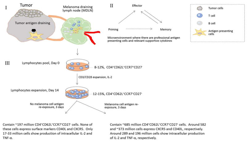
Figure 4.
Schematic Figure of the 14-day expansion of MDLN samples from Stage III melanoma patients. I. Cells within MDLN sample have experienced tumor antigens. I and II. In MDLN microenvironment where there are functional antigen presenting cells and supportive cytokines, T cells can undergo priming, effector and memory phase. III. The ex vivo activation by CD3/CD28 beads and expansion by IL-2 resulted in a kinetic growth of CD4+ TCM subset with negative CD27 expression (CD4+CD62L+CCR7+CD27−). Although the subsets do not proliferate or exhibit functional property in cultures with no antigen re-stimulation, the subsets exhibited a dramatic increase of total cell number in response to melanoma cell antigen re-exposure. The subsets also exhibited dramatically increased surface expression of CD40L and CXCR5 and intracellular production of IL-2 and TNF-α.

