Figure 2.

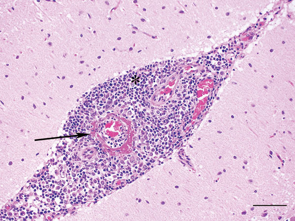
Cerebellum of dog infected with Hendra virus, showing expansion of the meninges with inflammatory infiltrates (*) and marked vasculitis (arrow). Scale bar indicates 75 μm.

Cerebellum of dog infected with Hendra virus, showing expansion of the meninges with inflammatory infiltrates (*) and marked vasculitis (arrow). Scale bar indicates 75 μm.