
Figure 2.
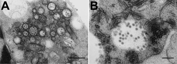
Electron micrographs of Middelburg virus isolate SAE25/2011 in baby hamster kidney cell culture. A) Several enveloped virions consisting of a dense core and surrounded by a translucent layer are shown in the vesiculated endoplasmic reticulum. The virus has elongated forms and numerous precursor nucleocapsids in the cytoplasm. Many of the nucleocapsids are associated with the outer surfaces of the vesiculated endoplasmic reticulum. Scale bar indicates 500 nm. B) Virions in a cytopathic vacuole are surrounded by elongated forms of the virus. Scale bar indicates 200 nm. Micrographs courtesy of Stephanie van Niekerk et al.