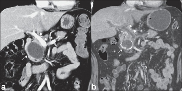
Figure 3.

Portal vein thrombosis after EUS-guided pancreatic cyst ablation.[20] (a) Initial CT scan: A 5.2 cm cyst (arrow) located adjacent to the main portal vein (arrow heads) at the head of the pancreas. (b) Follow-up CT scan after second cyst ablation: Cyst (arrow) with rim calcification and portal vein thrombosis (arrow heads)
