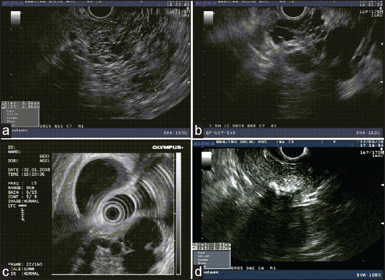
Figure 1.

The morphologic features on EUS imaging of the various subtypes of PCNs. (a) EUS feature of microcystic serous cystadenoma with honeycombing appearance in the neck of the pancreas in a 29-yearold female patient with abdominal pain, which was approximately 3.6 × 3.9 cm. (b) EUS image of a MCN in a 76-year-old male patient with presence of septations and approximately 3.7 × 2.8 cm in the head of the pancreas. (c) EUS image of an IPMN in a 78-year-old male with the papillary form. The diameter of the main pancreatic duct was 2.0 cm which was located in the head of the pancreas. (d) EUS appearance of the SPN in a 35-year-old female patient which presented a mixture echo mass about 1.42 × 0.95 cm and presence of calcifications in the head of the pancreas. PCN = Pancreatic cystic neoplasm, EUS = Endoscopic ultrasound, MCN = Mucinous cystic neoplasm, IPMN = Intraductal papillary mucinous neoplasm, SPN = Solid pseudopapillary neoplasm
