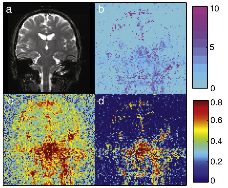
Fig. 7.
RetroICor BIC results for non-synchronized 3D bSSFP. a: Example image for anatomical reference. b: The number of BIC selected regressors calculated voxelwise. Inferior brain regions and regions of CSF show the largest number of selected voxels. c: the amount of variance reduction when the full set of 22 regressors is used for the regression and d: when the BIC determined model containing four regressors is used. Even with only a few regressors a large amount of variance can be removed.

