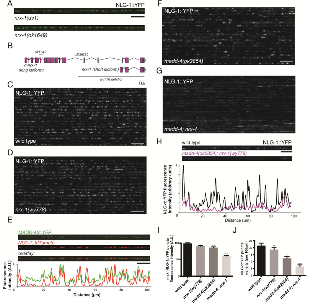
Figure 3. MADD-4 and NRX-1 are redundantly required for NLG-1 localization at the synapse.
(A) Confocal micrographs of nrx-1(ds1) and nrx-1(ok1649) mutant animals showing NLG-1::YFP clusters in the dorsal nerve cord. (B) Schematic representation of the nrx-1 locus, showing the α-neurexin and β-neurexin isoforms, the Mos transposon insertion site (ttTi26330) as well as the ok1649 and wy778 deletions. (C,D) Confocal micrographs showing the dorsal nerve cord of 20 wild-type (C) and nrx-1(wy778) (D) animals expressing NLG-1::YFP. (E) Confocal micrographs of neuronallysecreted MADD-4S::YFP and muscle NLG-1::dtTomato at the dorsal nerve cord. The fluorescence profile of MADD-4S::YFP (green) and NLG-1::tdTomato (red), normalized to their respective maximum intensity, are shown. (F,G) Confocal micrographs showing the dorsal nerve cord of 20 madd-4(ok2854) (F) and madd-4(ok2854); nrx-1(wy778) double (G) mutant animals. (H) The fluorescence profiles of NLG-1::YFP in a wild-type and madd-4(ok2854); nrx-1(wy778) double mutant animal are shown. Both fluorescence profiles were normalized to the wild-type maximum intensity. (I,J) Quantification of NLG-1::YFP puncta maximum fluorescence intensity (I) and synaptic density (J). An ANOVA and post hoc Tukey’s tests were used to determine significant differences between the different genotypes. Error bars indicate SEM. p values are *<0.05, ***<0.001, ****<0.0001. Scale bars: 5 µm (A,E), 10 µm (C,D,F–H).

