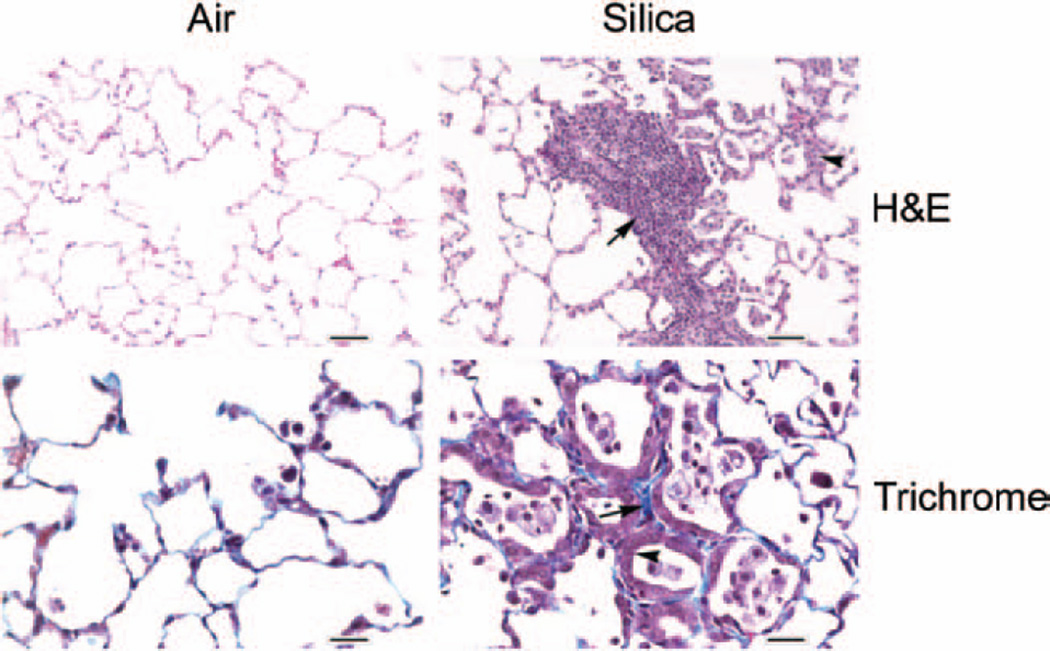
Figure 2.
Photomicrographs of lungs from control and silica-exposed rats. Lung samples obtained from control and crystalline silica-exposed rats were sectioned and stained with H & E (top panel) or Masson’s trichrome stain (bottom panel) as described in the text. The arrow and arrowhead in the H & E stained sections show type II pneumocyte hyperplasia and alveolar space filled with macrophages and neutrophils, respectively, in the silica-exposed rat lungs. The arrow and arrowhead in the trichrome stained sections show thickened alveolar septae and type II pneumocyte hyperplasia lining the alveolar septae, respectively, in the silica-exposed rat lungs Magnification: H & E = ×20; trichrome = ×40.

