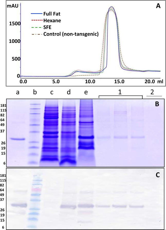
Fig. 3. Gel filtration of HBsAg.
(A) Gel filtration chromatogram for SFE-treated material (green line), hexane-treated (red line) , full fat material (blue line) and non-transgenic maize control (brown line). (B) SDS-PAGE of protein eluted from the gel filtration. Yeast-derived HBsAg was used as a positive control (lane a); protein standard marker (lane b); non-transgenic maize seed extract as negative control (lane c); transgenic maize seed extract (lane d); AS-purified HBsAg (lane e); fractions of protein eluted from gel filtration peak 1 (lane 1); protein eluted from gel filtration peak 2 (lane 2). (C) The corresponding anti-HBsAg western blot demonstrating the presence of HBsAg. The SDS PAGE and western blots presents data collected for SFE treated material and are representative of data collected for hexane treated and full fat material.

