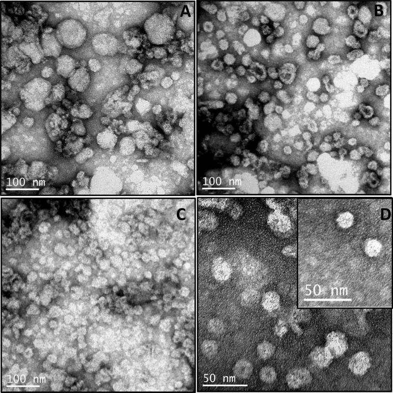
Fig.4. Electron microscopy of virus-like particles (VLPs).
TEM images of VLPs formed by HBsAg present in the void volume fractions of gel filtration of proteins obtained from (A) full fat germ; (B) hexane-treated germ; (C) SFE-treated germ; (D) VLPs of SFE-treated germ shown at higher magnification. (E) Elute obtained from peak 2 of the gel filtration chromatography (negative control). (F) Protein extract from non-transgenic maize material (negative control). (G) VLPs of yeast derived HBsAg (Meridian)(positive control). VLPs/proteins were negatively stained with 0.5% uranyl acetate and visualized by TEM.


