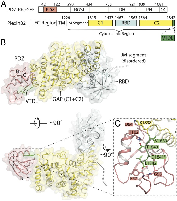
Fig. 1.
Crystal structure of the complex between PlexinB2cyto and the PDZ domain from PDZ–RhoGEF. (A) Domain structure of PlexinB2 and PDZ–RhoGEF. Residue numbers are based on human PDZ–RhoGEF and mouse PlexinB2, respectively. CC, coiled-coil; DH, Dbl-homology domain; EC-region, extracellular region; PH, pleckstrin-homology domain; RGSL, regulator of G protein signaling-like domain; TM, transmembrane region. The C1 and C2 segments in plexin together form the GAP domain. (B) Structure of the PlexinB2cyto/PDZ complex. The color scheme is the same as in A. N and C indicate the N and C termini of the PDZ domain, respectively. The dotted lines indicate the approximate location of the disordered JM-segment. (C) Interface between the PDZ domain and the VTDL motif in PlexinB2. *The side chain of D1841 is not built because of poor density.

