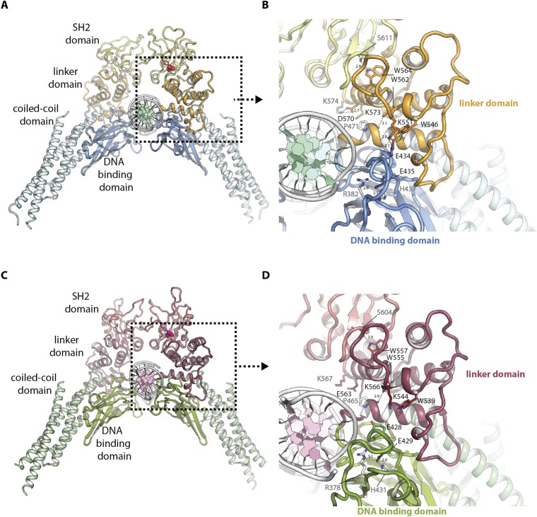
Fig. S1.
Functional architecture of mouse STAT3 and human STAT1 in complex with DNA. (A) Functional architecture of mouse STAT3 in complex with DNA (PDB ID code 1bg1) with coiled-coil domain (light blue), DNA-binding domain (blue), linker domain (orange), SH2 domain (yellow), and DNA (white and green). Phosphotyrosine is highlighted in red. (B) Detailed view of A showing the interface between mouse STAT3 and DNA. Side chains of residues mutated in this study are displayed as colored sticks with black type, residues interacting with them are displayed as white sticks with gray type. Hydrogen bonds are highlighted by yellow dashes and distances labeled in angstroms. (C) Functional architecture of human STAT1 in complex with DNA (PDB ID code 1bf5) with coiled-coil domain (light green), DNA binding domain (green), linker domain (mauve), SH2 domain (light mauve), and DNA (white and light pink). Phosphotyrosine is highlighted in pink. (D) Detailed view of C showing the interface between human STAT1 and DNA. Side chains of residues equivalent to those mutated in mouse STAT3 in this study are displayed as colored sticks with black type, residues interacting with them are displayed as white sticks with gray type. Hydrogen bonds are highlighted by yellow dashes and distances labeled in angstroms.

