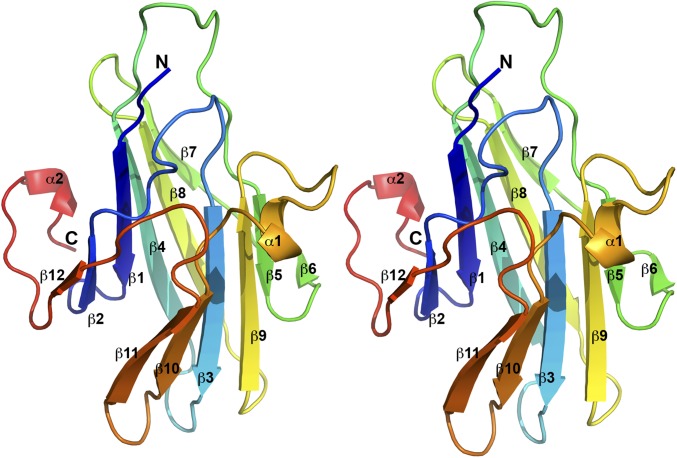
Fig. 1.
The structure of VACV C7 displays a previously unidentified fold. A stereo view of the C7 structure is depicted. The secondary structures are labeled and shown in rainbow color with the N terminus in blue and the C terminus in red. The structure of C7 consists mainly of two curved layers, each comprised of a six-stranded antiparallel β sheet. Structure homology search did not find any match, suggesting that C7 adopts a novel fold.

