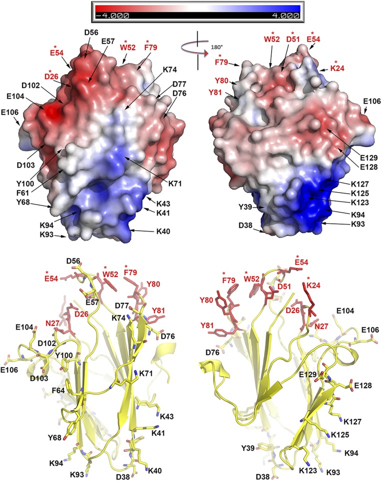
Fig. 2.
The functional site for C7 centers at three spatially clustered loops. (Upper) Electropotential surface of C7 shown in two views with 180° rotation. The residues that were mutated in this study are indicated by arrows. The residues whose mutations had no effect on viral replication are labeled in black. The residues whose mutations abolished viral replication are indicated in red; those that are conserved in C7-equivalent homologs are marked with red asterisks. (Lower) C7 is shown in a cartoon presentation with the secondary structures. The coloring scheme is the same as above. Notice that the key functional residues are clustered on the top of the β sandwich. The three functional loops form a unique molecular claw.

