Fig. 1.
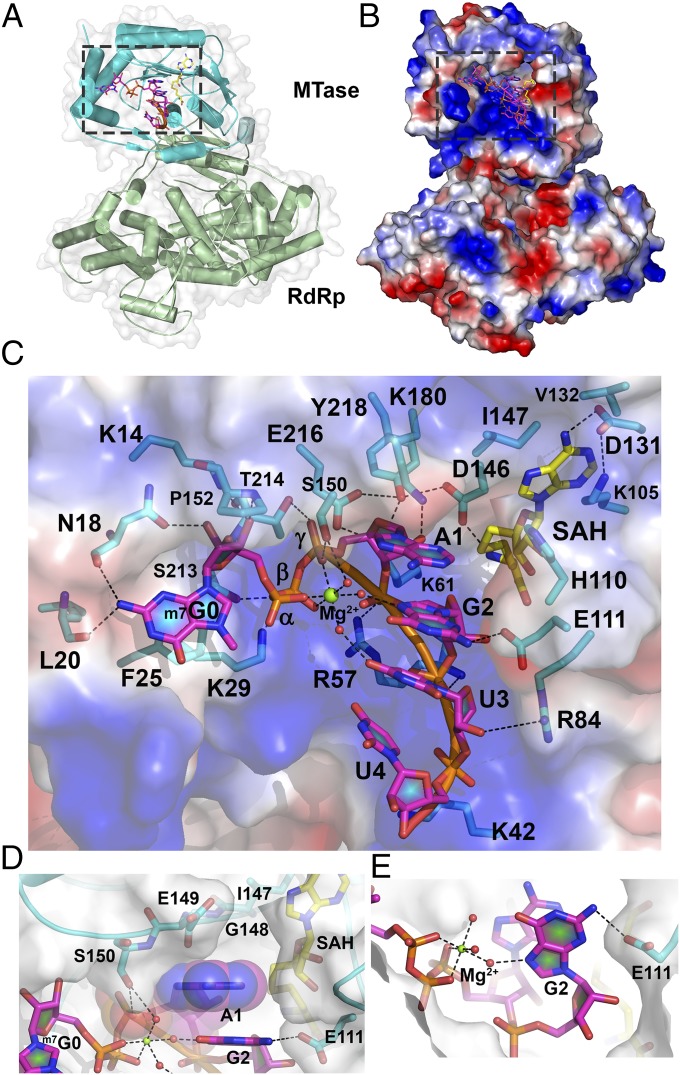
Structure of the ternary complex between DENV3 NS5, capped RNA, and SAH. (A) Crystal structure of NS56–896 (MTase domain, cyan; RdRp domain, green) bound to RNA (5′-m7G0pppA1G2U3U4GUU-3′; magenta sticks) and SAH (yellow sticks). The RNA/SAH binding site is boxed. (B) Electrostatic surface representation of NS5 (positive charges are in blue, and negative charges are in red). Capped RNA (magenta) and SAH (yellow) are shown as sticks. (C) Close-up view of the RNA and SAH binding sites as boxed in B. Protein residues binding RNA or SAH are represented as sticks (cyan) and labeled. Capped RNA nucleotides are labeled. Dashed lines indicate polar interactions. Mg2+ is displayed as a green sphere, and water molecules are shown as red spheres. (D) Interactions established between adenine A1 from the bound RNA and NS5, illustrating the tight shape complementarity with adenine only. (E) Hydrogen bonds formed between E111, the capped RNA second nucleotide G2, and the bound Mg2+ ion.

