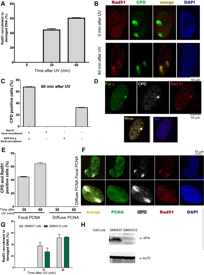
Fig. S7.
Rad51 and polη are recruited to CDPs in cells transiting S phase. (A) U2OS cells were UV-irradiated using polycarbonate filters and fixed at the indicated time points. Percentages of CPD-positive nuclei with Rad51 enrichment at CPD spots were calculated in 100 CPD-positive nuclei. Average values were calculated by comparing three independent experiments. (B) Representative nucleus showing Rad51 recruitment to CPDs. (C) U2OS cells were transfected with GFP-polη. Twenty-four hours later, samples were UV-irradiated using polycarbonate filters and fixed at 60 min post-UV irradiation. CPDs and Rad51 were detected using specific antibodies, and GFP autofluorescence was used to detect polη. The percentage of double-positive (Rad51 and polη), single-positive (Rad51 or polη), and double-negative (neither rad51 nor polη) recruitment to CPDs was calculated after analyzing 100 CPD-positive nuclei. Average values were calculated by comparing two independent experiments. (D) Representative nucleus showing Rad51 and Polη simultaneous recruitment to CPDs. (E) U2OS cells were transfected with GFP-proliferating cell nuclear antigen (PCNA). Twenty-four hours later, samples were UV-irradiated using polycarbonate filters and fixed 30 and 60 min later. CPDs and Rad51 were detected using specific antibodies, and GFP autofluorescence was used to detect PCNA. The percentages of Rad51 recruitment to CPDs in S phase (GFP-PCNA focal) and outside S phase (pannuclear GFP-PCNA) were calculated after analyzing 100 CPD-positive nuclei transfected with GFP-PCNA. Average values were calculated by comparing two independent experiments. (F) Representative nuclei showing Rad51 recruitment to CPDs in cells in S phase (GFP-PCNA focal) and the lack of Rad51 localization in CPDs accumulated outside S phase (pannuclear GFP-PCNA). (G) GM0637 (XPA-proficient) and GM04312 (XPA-deficient) fibroblasts were UV-irradiated using polycarbonate filters. At the indicate time points, samples were fixed and subjected to immune detection of Rad51 and CPDs. Percentages of CPD-positive nuclei with Rad51 enrichment at CPD spots were calculated in 100 CPD-positive nuclei. Average values were calculated by comparing two independent experiments. (H) Western blot showing XPA levels in GM0637 and GM04312 fibroblasts.

