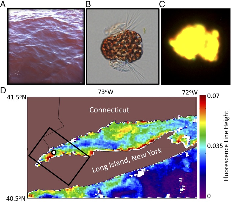
Fig. 1.
(A) Photograph of red water in WLIS during the observed M. rubrum bloom. Photo courtesy of Kay Howard-Strobel (University of Connecticut, Groton, CT). (B) Micrograph of the ciliate M. rubrum with enslaved cryptophyte chloroplasts. Micrograph courtesy of National Oceanic and Atmospheric Administration Phytoplankton Monitoring Network. (C) Fluorescent microscopy under green light excitation showing characteristic yellow fluorescence (565–570 nm) (26) of M. rubrum due to high amounts of phycoerythrin pigment. (D) Satellite image of Chl a fluorescence line height from the MODIS Terra sensor on September 23, 2012 shows a dense bloom in WLIS, where M. rubrum was sampled at 1 × 10⁶ cells per liter (black circle). The black rectangle outlines the swath of the coincident HICO imagery obtained from the International Space Station.

