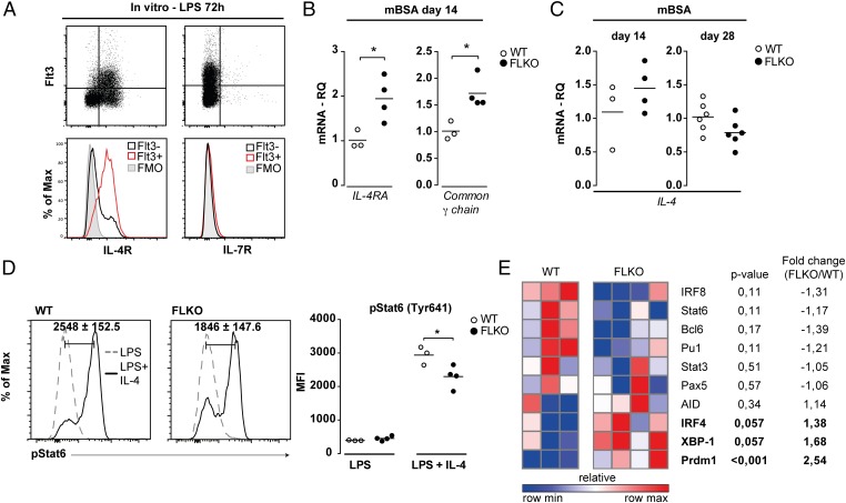
Fig. 6.
Deficiency of functional Flt3 signaling in activated B cells results in impaired IL-4–induced activation of Stat6. (A) Expression of the IL-4R and IL-7R on activated Flt3+ WT B cells (gated on CD19+ cells) after 72 h of LPS stimulation. (B) mRNA for IL-4R and the common γ-chain in the spleen of FLKO and WT mice. (C) IL-4 mRNA in the spleen of WT and FLKO mice. (D) Phosphorylation of Stat6 (Tyr641) in WT and FLKO B cells after 72 h stimulation with LPS (dashed line) or LPS + IL-4 (solid line). Cells are gated on B220+ lymphocytes. Histograms represent the difference between LPS and LPS + IL-4 simulated cells. (E) Gene expression heat map of spleen cells from FLKO and WT mice at day 3 after in vitro LPS + IL-4 stimulation. Gene expression of target genes was normalized against GAPDH- and LPS-stimulated cultures. *P < 0.05. Statistical analysis was performed using the unpaired Student’s t test.

