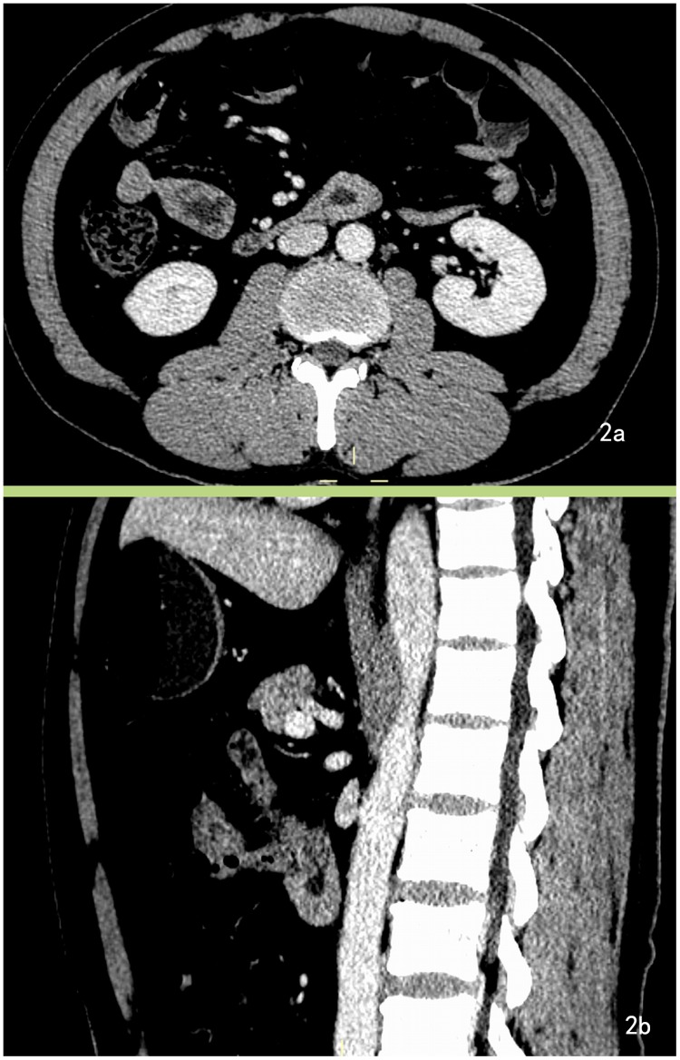
Fig 2. GIST overlooked in CT images.
A 64-year-old male with duodenal GISTs presented with melena for the past 20 days. A. Enhanced CT image demonstrates bowel wall thickening at the third segment on the axial section of the CT image and was overlooked. B. Post-procedural CT images (sagittal section) reveal the lesion more clearly. One radiologist had overlooked the lesion, while the other radiologist had misdiagnosed it as a heterotopic pancreas.

