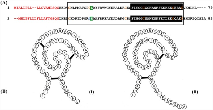
Fig 1. Amino acid sequences of EgKI-1 (1) and EgKI-2 (2).
A. Signal sequences (18 amino acids) are in red, the Kunitz domain of both proteins is boxed and the Kunitz family signature highlighted in black. The conserved cysteine residues are shown in orange; EgKI-1 has six whereas EgKI-2 has five with one position replaced by a glycine (blue). The P1 reactive sites of both proteins are highlighted in green. B. Schematic diagram of (i) EgKI-1 showing three disulphide bridges, and (ii) EgKI-2 presenting two disulphide bridges.

