
Fig 6.
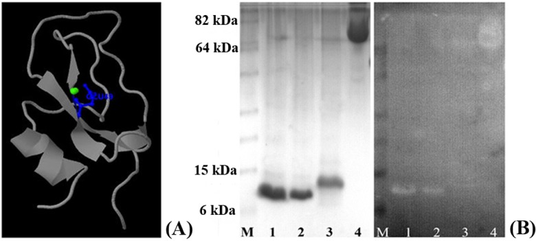
(A) Predicted calcium binding site of EgKI-1, shown in blue. (B) Calcium binding assay: SDS-PAGE gel (left) and corresponding western blot membrane of the calcium binding assay showing clear bands corresponding to the EgKI-1 protein: M-Marker; 1, 6 μg EgKI-1; 2, 2 μg EgKI-1; 3, 6 μg EgKI-2; 4, 6 μg BSA.