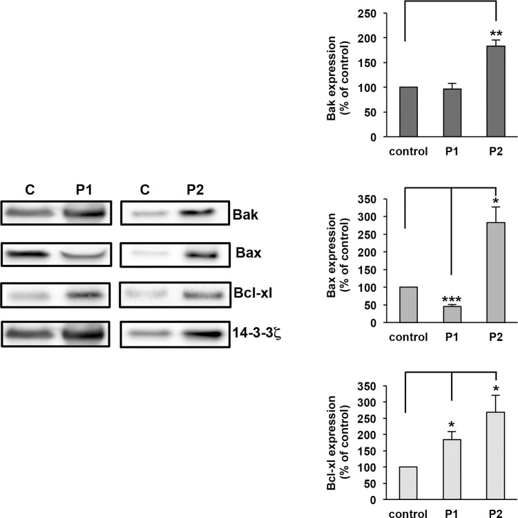
Fig 1. Expression of apoptotic proteins in patients with VWD-type 2B.
Washed platelets (2.5 x10⁸ platelets/mL) from controls (C) or P1 and P2 were lysed and then equal numbers of platelets were loaded. Apoptotic proteins were assessed by immunoblotting with anti-Bak, anti-Bax, anti Bcl-xL and anti-14-3-3ζ antibodies. Data are expressed as the ratio of apoptotic protein expression versus 14-3-3ζ expression. Then, the ratio of an apoptotic protein for P1 or P2 was compared with the corresponding ratio for control (100%). Results are means ± Standard Error of the Mean (SEM) from three independent experiments. *p = 2.8.0x10-2, **p = 2.3x10⁻³, ***p = 2.9x10⁻⁴.

