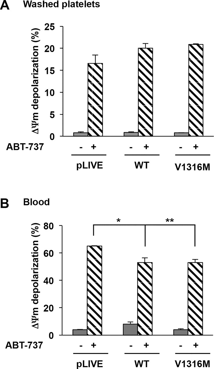
Fig 6. ΔΨm depolarization in mice expressing VWD-type 2B.
ΔΨm depolarization was determined in washed platelets pretreated for 30 minutes with TMRE-PE (500 nM final concentration) at 37°C. Then platelets were incubated further for 60 minutes with or without ABT-737 (10 μM final concentration) and analyzed by flow cytometry. Results are expressed as a percentage of depolarized cells. Means ± SEM from three independent experiments are shown. *p = 2,5 x 10⁻² and **p = 5.0 x10⁻³ (unpaired Student t test).

