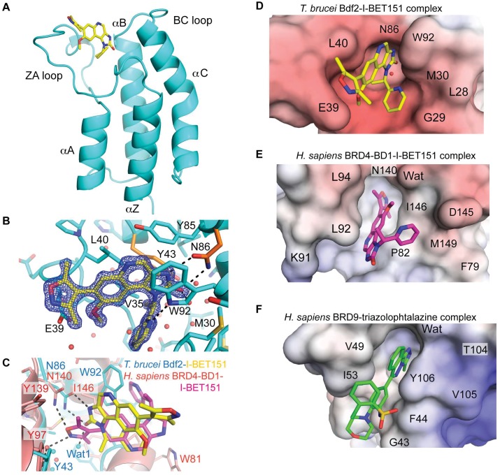
Fig 7. Structural analysis of I-BET151 binding to Bdf2.
(A) Structure of the Bdf2 bromodomain in complex with I-BET151 (PDB code 4PKL). (B) Electron density of I-BET151 and key residues for ligand recognition. The two conserved residues mutated to alanine in the Bdf2 mutant are colored in orange. (C) Superimposition of Bdf2 (cyan) in complex with I-BET151 (yellow) onto human BRD4-BD1 (light red, 3ZYU) in complex with I-BET151 (magenta). (D to F) Electrostatic surface potential shown between -10 kT/e (red) and +10kT/e (blue) of (D) trypanosome Bdf2 bromodomain in complex with I-BET151, (E) human BRD4-BD1 in complex with I-BET151 (3ZYU), and (F) human BRD9 in complex with a triazolophtalazine inhibitor (4NQN).

