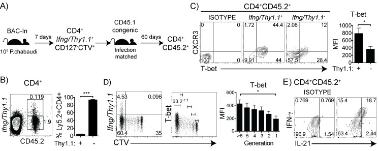
Fig 8. IFN-γ+ memory cells co-produce IL-21, and Ifng-accessibility and T-bet are maintained by division during chronic malaria infection.
(A) BAC-In mice (CD45.2) were infected and at day 7 post-infection, CD4+ Ifng/Thy1.1 + T cells were purified, CTV labeled, and adoptively transferred into infection-matched CD45.1 recipients. At day 60 post-infection, splenocytes were analyzed by flow cytometry. (B) Dot plot and bar graph shows expression of Ifng/Thy1.1 in recovered donor cells. (C) Contour plots gated on CD4+CD45.2+ show expression of CXCR3 and T-bet in recovered Ifng/Thy1.1 + and Ifng/Thy1.1 - populations. Bar graph shows T-bet MFI. (D) Ifng/Thy1.1 and T-bet within the CTV- maximally divided cells. Bar graph shows T-bet MFI in each division (E) IFN-γ and IL-21 expression ex vivo in CD4+CD45.2+ donor cells. Data are representative of two independent experiments with 3–4 mice per experiment. Statistical significance shown using Students t-test. Error bars represent SEM; ★ p < 0.05, ★★★ p < 0.001.

