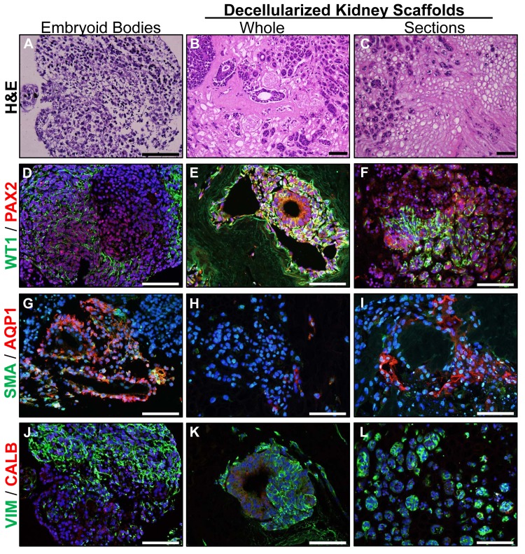
Fig 3. Renal developmental markers are upregulated by renal ECM.
hESC were differentiated as embryoid bodies (A) or cultured in whole kidneys (B) or sections of kidneys (C) where cells were typically observed in the medulla and medullary rays. (D-F) Renal developmental markers WT1 and PAX2 were upregulated in whole or sections of decellularized kidneys when compared with embryoid body differentiation. (G-I) AQP1, a marker of proximal tubules, was expressed in tubule-like structures in embryoid bodies and kidney sections, but not in whole kidneys. (J-L) Vimentin, a mesenchymal and mesangial marker, was expressed under all culture conditions. Other markers of mature renal cell types including SMA (mesangial and vascular smooth muscle marker) and Calbindin (renal distal tubules) were not expressed. Nuclei were visualized with DAPI (blue); scale bars = 100 μm.

