
Figure 5. The detection of surface-bound TrpRS on INF-γ-treated OSCC cells.
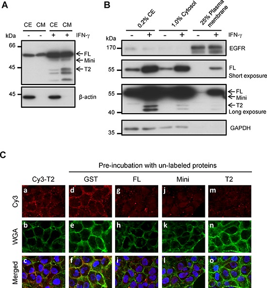
A. The detection of intracellular and extracellular TrpRS expressions in IFN-γ-treated OSCC cells. OEC-M1 cells were treated with IFN-γ (200 U/ml) for 24 h. Cell extracts (CE) and CM were obtained from control and IFN-γ treated cells, and the proteins were detected via Western blot using an anti-TrpRS antibody. β-actin was used as the loading control. B. TrpRS was detected in the plasma membrane (PM) fraction of IFN-γ-treated OEC-M1 cells. OEC-M1 cells were treated with IFN-γ (200 U/ml) for 24 h. The whole-cell extract (CE), cytosolic (Cytosol) and PM fractions were prepared as described in the Materials and Methods section. The proteins were subjected to Western blot using anti-EGFR, anti-TrpRS and anti-GAPDH antibodies as indicated. C. Cy3-labeled T2-TrpRS was detected on the cell surface via immunofluorescence staining as described in the Materials and Methods section. Cells pre-incubated without a–c. or with 50 μg/ml unlabeled GST d–f. full-length GST-TrpRS g–i. GST-mini-TrpRS j–l. or GST-T2-TrpRS m–o. are presented. The Cy3-labeled T2-TrpRS (red), WGA (green) and merged images are presented as indicated. DNA was stained with Hoechst 33258 (blue).