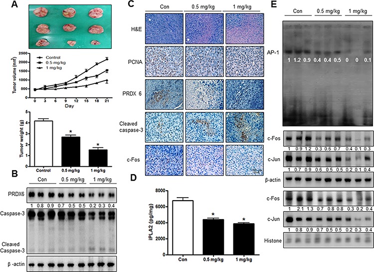
Figure 6. SVT inhibited tumor growth in vivo xenograft.

Tumor volumes, weights, and images of normal mice A. The expression of PRDX6 and Caspase-3 was detected by western blotting B. β-actin protein was used an internal control. Tumor sections of mice were analyzed by H&E, PCNA, PRDX6, Caspase-3 and c-Fos by immunohistochemistry C. Expression of iPLA2 was detected by ELISA kit D. AP-1 activity in tumor tissue E. The resultant tissues were developed with DAB, and counterstained with hematoxylin. Scale bar indicates 50 mm. *(P ≤ 0.05) indicates statistically significant differences from control cells.
