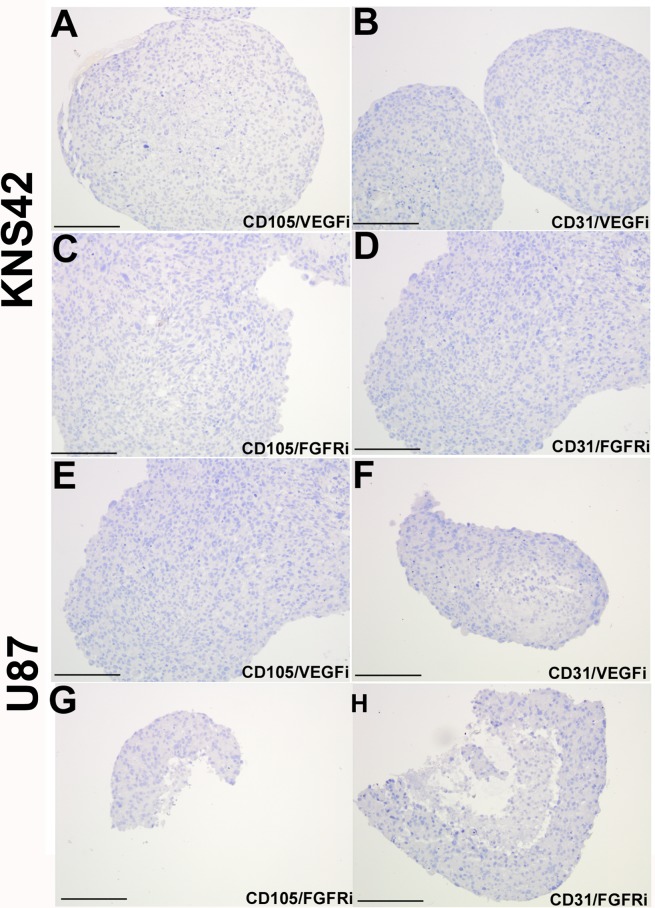
Figure 6. Loss of tumor-derived endothelial marker expression upon VEGF or FGFR inhibition in vitro.
KNS42 and U87 aggregates were cultured for 7 days in the RCCS and subsequently exposed to the CBO-P11 VEGF inhibitor (10 μM) or PD16686 FGFR inhibitor (15 μM) respectively for 3 days. A.–D. Absence of CD105 and CD31 staining upon exposure to either inhibitor in KNS42 cells. E.–H. Absence of CD105 and CD31 staining upon exposure to either inhibitor in U87 cells. Scale bar A-H = 200μm. Whole field views of aggregates are shown in all cases to indicate complete absence of endothelial marker staining.

