
Figure 6. PYK2 affects EGF-induced IL8 expression while IL8 induces PYK2 phosphorylation.
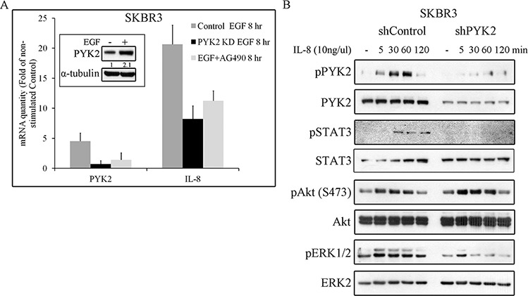
A. EGF-induced IL8 expression in STAT3 and PYK2-dependent manner. The mRNA levels of PYK2 and IL8 in the control, AG490-treated (10 μM) and PYK2-depleted SKBR3 cells were assessed in response to EGF (50 ng/ml, 8 hr) stimulation using real-time PCR. The mRNA levels are presented as fold of un-stimulated control cells. The mean values ± s.d. of three experiments are shown. An insert of PYK2 protein levels in control serum-starved and EGF-stimulated cells is shown. Quantification was done as in Figure 5D B. IL8 induces phosphorylation of PYK2 and activates downstream pathways. Control and PYK2-depleted SKBR3 cells were serum-starved for 24 hr and then stimulated with IL8 (10 ng/ml) for the indicated time periods. Total cell lysates were prepared and analyzed for the activation of different signaling pathways using WB analysis and phospho-specific antibodies as indicated. Reproducible results were obtained in three independent experiments.