
Figure 1. Identification by immunohistochemistry (IHC) of STMN1 and its multiple phosphor-sites in the primary tumor and Kaplan-Meier analysis of DFS in breast cancer patients with high or low STMN1 expression and the expression of its multiple serine phospho-sites.
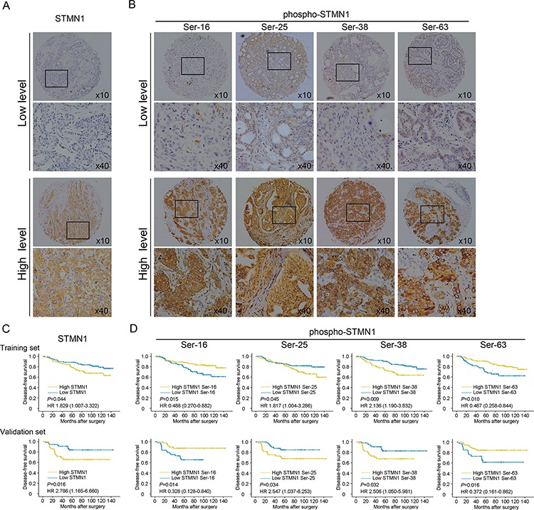
A. Representative IHC staining of high and low expression of STMN1 in the large (400×) and small images (100×). B. Representative IHC staining of high and low expression of multiple phosphor-sites (Ser-16, Ser-25, Ser38, Ser63) in the large (400×) and small images (100×). C. Kaplan-Meier analysis of DFS in the training set. D. Kaplan-Meier analysis of DFS in the validation set.