
Figure 8. Panicein A hydroquinone presents a strong docking cluster close to the doxorubicin binding site in AcrB structure and in Patched structural model.
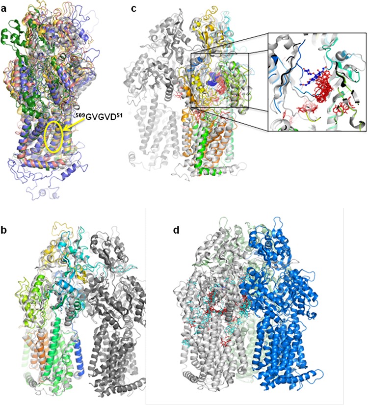
a. Superposition of the different models obtained for human Patched. The transmembrane domain is consistent between the different results, especially the residues GVGVD taking part of the proton transfer. b. Monomer of the final selected Patched model superposed on the AcrB structure. The trimer of AcrB is colored in three different greys, the Patched model is in rainbow color. c. Panicein A hydroquinone / AcrB docking results. All the panicein A hydroquinone possible positions are presented. The zoom corresponds to an enlargement of the dxr binding pocket. The proteins structures are colored as in (b), dxr is in blue, panicein A hydroquinone is in red. d. AcrB docking results of panicein C (light blue) and panicein A hydroquinone (red) presented on a monomer of the AcrB structure. The three monomers are colored in light grey, light green, and blue.