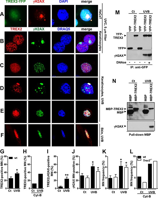
Figure 9. TREX2 co-localizes and interacts with γH2AX in micronuclei.

A. HaCaT cells expressing exogenous YFP-TREX2 and B. primary keratinocytes UVC-irradiated (100 J/m2) through 5-μm micropore filters, C. keratinocytes globally-irradiated with UVB (150 J/m2), D. containing micronuclei or E. detaching, and F. UVB-treated skin samples were stained with γH2AX and TREX2 antibodies, as indicated. G. Percentage of TREX2 single-positive micronuclei scored in the absence and H. in the presence of Cyt-B, and I. micronuclei double-positive for TREX2 and γH2AX or J. single-positive for γH2AX. K. Micronuclei frequency in untreated or UVB-irradiated (150 J/m2) wt and Trex2−/− keratinocytes in the absence or L. presence of Cyt-B. In the presence of Cyt-B, only binucleated cells were scored. The data represent the mean and SEM of three independent experiments (G, I, J, K), and the mean of one (H, L) analysis of up to 1, 000 cells. M. TREX2 interacts with γH2AX as shown by coimmunoprecipitation of γH2AX and YFP-TREX2 after UVB (150 J/m2) irradiation of HaCaT cells stably expressing YFP-TREX2 and by N. MBP pull-down assays. Nuclei were counterstained with DRAQ5 or DAPI, as indicated. Original magnification was 100x (A) and 63x (B-F) P values between untreated and UVB-treated keratinocytes calculated by unpaired Student's t test: *P < 0.05, **P < 0.01.
